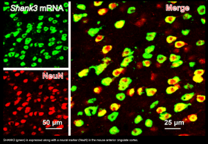
Neuroscientists from MIT and colleagues in China have identified a brain region, the anterior cingulate cortex (ACC), that is linked to altered social interactions in a mouse model of autism. The study, published in Nature Neuroscience, found that restoring activity in the ACC reversed social traits associated with autism in the mouse model. The researchers focused on the ACC, known for its role in social functions, after previous studies failed to definitively link the prefrontal cortex (PFC) to altered social interactions in mice with mutations of the SHANK3 gene associated with autism spectrum disorder (ASD).
Mice with SHANK3 mutations exhibited avoidance of social interactions, and the study showed that SHANK3 knockout mice had structural and functional disruptions in synapses between excitatory neurons in the ACC. The loss of SHANK3 in excitatory ACC neurons alone disrupted communication and decreased activity during social tasks. However, when the ACC neurons were activated in SHANK3 mutant mice using optogenetics and specific drugs, social behavior improved.
The researchers plan to further investigate downstream brain regions that modulate social behavior and aim to understand social deficits in neurodevelopmental disorders. The findings are promising as anatomical structures in the ACC were found to be altered or dysfunctional in individuals with ASD, indicating potential relevance to the SHANK3 gene findings. The research was funded in part by the Natural Science Foundation of China, with Guoping Feng being supported by various institutions including the National Institute of Mental Health and the Hock E. Tan and K. Lisa Yang Center for Autism Research at MIT.
